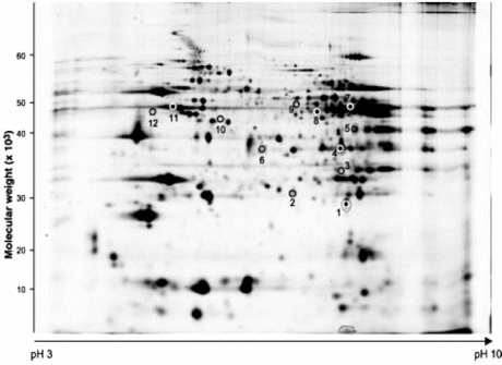
Fig. (1).
2D gel electrophoresis reveals proteins that were differentially expressed in PLM KO mice relative to wild-type controls. Spots of interest were excised and subjected to mass spectrometry analysis. Used with permission: Bell et al., Characterization of the phospholemman knockout mouse heart: depressed left ventricular function with increased Na-K-ATPase activity. Am J Physiol Heart Circ Physiol 2008; 294: H613-H621.

