
Figure 3. Determination of glial and neuronal markers (GFAP & NSE, respectively) at different time points of the culture by western immunoblotting.
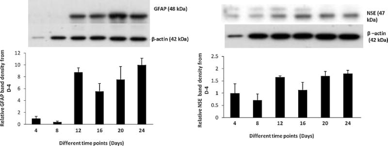
GFAP protein signal was found to be low at the initial two time points and rapidly increased to several folds at day 12 and stayed elevated in the remaining days of the culture. NSE signal was strong at day 4, weak at day 8 and increasing rapidly at day 12. The signal remains elevated in the remaining time points of the culture.