
Figure 6. Detection of APP protein in neuronal cultures at different time points.
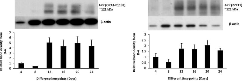
Levels of APP were detected by western blot from the cell lysate as described in the text. The result is consistent with the other findings, i.e., total APP levels are increased several fold at day 12 and stay elevated thereafter. Two distinct isoforms of APP (the top band at 128 kDa represents the mature/glycosylated and 121 kDa band represents immature/ deglycosylated form) was detected.