
Figure 9. DNA transfection of neuronal culture and visualization by fluorescence microscopy.
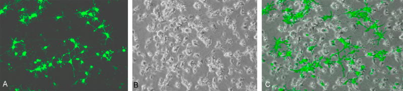
Cells were transfected with pmaxGFP at day 12. Transfection was carried out by lipid based transfection reagent (Transfectin®; Bio-Rad). Transfected cells started expressing ‘green fluorescent protein’ approximately after 36 hours of transfection and maximum expression was noticed after 48 hours. Percentage calculation after manual counting of green transfected cells and non-transfected cells revealed an approximate transfection efficiency of 25-30%.