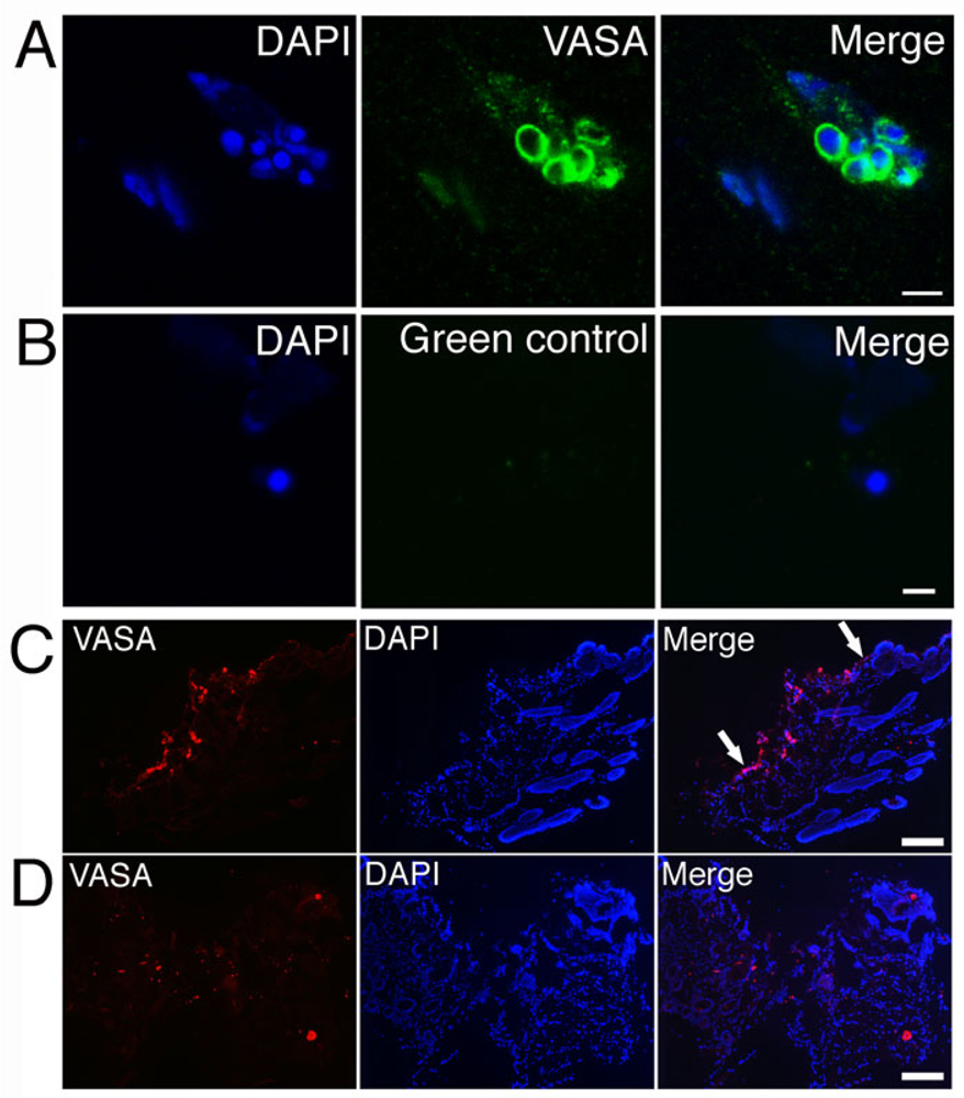
Figure 5.
VASA-expressing Dot cells regenerate epithelial cells. VASA expression on Dot cells was induced after 10 day co-cultured with dermal fibroblasts. 5A shows a group of Dot cells that express VASA on their surface. The control section in 5B shows no positive stain. Figure 5C shows a 3-day post-wound section of Dot cell transplanted mouse with strong VASA-expressing cells along the epidermal layer (between arrows in 5C). 5D shows VASA expression in the saline control mouse wound. Bar in 5A = 5 µm; bar in 5B = 2 µm; bars in 5C and 5D = 200 µm.

