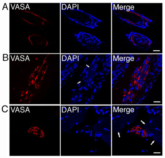
Figure 7.
Dot cells regenerate epithelial and dermal cells. One million Dot cells were transplanted via tail vein into skin-wounded adult mice. Wounds were stained using rabbit anti-VASA antibody and followed with fluorescent-labeled secondary antibody. VASA expression was examined. Confocal images indicate that the expression of VASA is found in hair follicles (7A), in the dermal wound area near by, but not related to, cell nuclei (arrows in 7B), and in the area that was surrounded by smooth muscle fibers (arrows in 7C). Bars = 20 µm.

