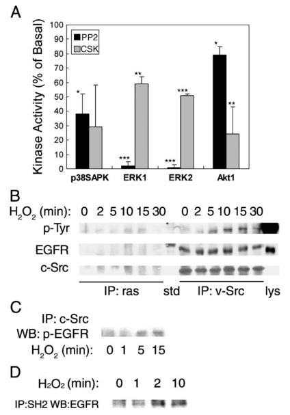
Fig. 4.
Assessment of the role of Src. (A) Cells pretreated with 5 μM PP2 for 30 min or transfected with Csk plasmid were stimulated with 0.5 mM H2O2 for 5 min. Kinase activities were determined as described in Fig. 1 legend (*p < 0.05, **p < 0.01, ***p < 0.001). (B) Cells were lysed following treatment with 0.5 mM H2O2 for the time period indicated. One milligram total protein was immunoprecipitated with either anti-ras (left) or anti-v-Src (right) monoclonal antibody followed by blotting with (a) anti-phosphotyrosine antibody (shown are bands that resolve at 180 kDa); (b) anti-EGF receptor antibody; and (c) anti-c-Src antibody; std, molecular weight standard; Lys, lysate of EGF-treated A431 cell as positive control. (C) Cells were lysed following treatment with 0.5 mM H2O2 for 0-15 min. Lysates containing 1 mg total protein were immunoprecipitated with anti-c-Src goat antibody followed by blotting with anti-phospho-EGFR monoclonal antibody. (D) Association of the SH2 domain of Src with the EGF receptor. Cells were lysed following treatment with 0.5 mM H2O2 for the time period indicated. Cell lysates were mixed with GST-SH2 fusion protein conjugated with agarose beads. The pull-down products were resolved by SDS-PAGE and immunoblotted with anti-EGF receptor antibody.

