FIGURE 2.
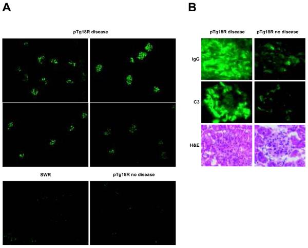
Kidney pathology in autoimmune pTg18R mice. A, kidney sections stained with goat anti-mouse IgG-FITC at a low magnification (10x). B, IgG and complement factor 3 (C3) deposition revealed by immunofluorescence, and glomerular inflammation revealed by H&E staining for representative diseased (left panels) and non-diseased (right panels) pTg18R mice.

