
Fig. 1.
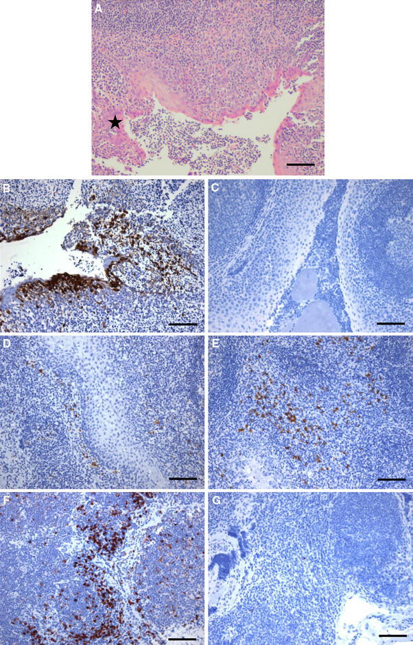
Tonsillitis in macaques infected with H5N1 virus. a Epithelial necrosis and loss (star) and infiltration of neutrophils amongst the normal lymphoid component of the tonsil and in the lumen. HE, scale bar 400 μm. b Virus infection in tonsillar epithelial cells, infiltrating leukocytes and underlying lymphoid tissue demonstrated by immunohistochemistry. c Absence of virus-antigen-positive cells in an A/Texas-infected animal (this image is representative of all groups other than H5N1). d Loss of CD83 positive cells 4 days pi with H5N1 virus. e Normal levels of CD83-positive dendritic cells in control tonsil, and representative for all other groups. f Pronounced apoptosis of cells, detected by IHC for activated caspase 3, in tonsil 4 days pi with H5N1 virus. g No apparent apoptosis in tonsils from any of the other groups. (b–g) IHC; scale bars 300 μm