
Fig. 3.
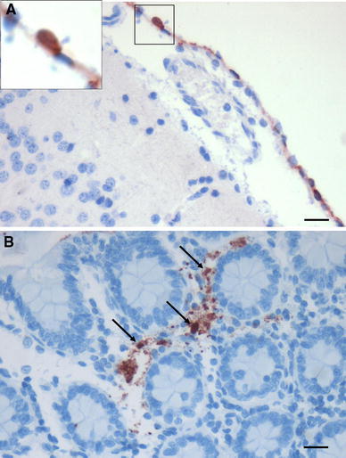
Detection of influenza virus antigen in extrapulmonary tissues 2–4 days pi in H5N1-infected animals. a Viral antigen in the mesothelial cells of the meninges of a macaque infected with H5N1 virus 4 days earlier. Inset higher magnification of a virus-antigen-positive cell, boxed in the main image, to illustrate the nuclear labeling. b Nuclear labeling (arrows) for H5N1 virus in cells in the lamina propria of the colon of a macaque terminated on day 2 pi. The granular signal surrounding the positive nuclei may be cellular debris from infected cells or some cytoplasmic staining. a, b IHC, scale bars 20 μm