
Fig. 5.
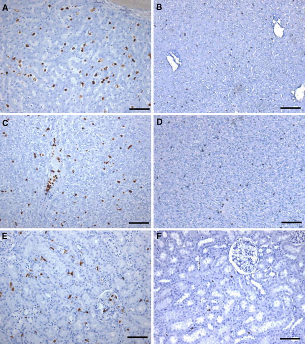
a, c, e Immunohistochemical detection of myeloid lineage cells and HIF-1α expression in liver and kidney of H5N1-virus-infected macaques compared to the other four groups. The microphotos are representative of expression throughout days 1–7 pi. a IHC for the myeloid cell marker Mac387 in liver reveals an increase in both numbers and size of macrophage-like cells including Kupffer cells on day 4 pi. c HIF-1α expression in intravascular and intrasinusoidal leukocytes in the liver of a H5N1-virus-infected macaque on day 1 pi. e Increased numbers of interstitial and intravascular Mac387-positive cells in the kidney of an H5N1 virus-infected animal, day 7 pi. b, d, f For each tissue and marker, similarly stained sections from a sham-infected animal and also representative of the other three virus-infected groups. Scale bars 200 μm