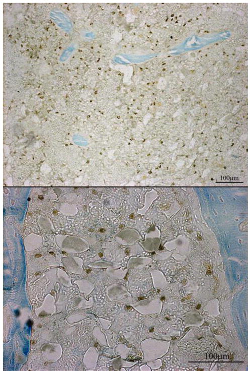
Figure 9. GFP Positive Cells in the Marrow.

GFP positive cells were found in the marrow 48 days after surgery. At all of the time points there was evidence of some GFP positive staining, but it was not until day 48 that there were large populations of GFP positive cells in the marrow spaces. Group C48 showed evidence of a smaller population of GFP positive cells in comparison to the other groups euthanized on day 48 (A48, B48, and D48). Cells were also present in other locations (cortices and pin sites), but the most consistent location for the MSC populations was in the medullary marrow (top) and the marrow within the periosteal callus (bottom).
