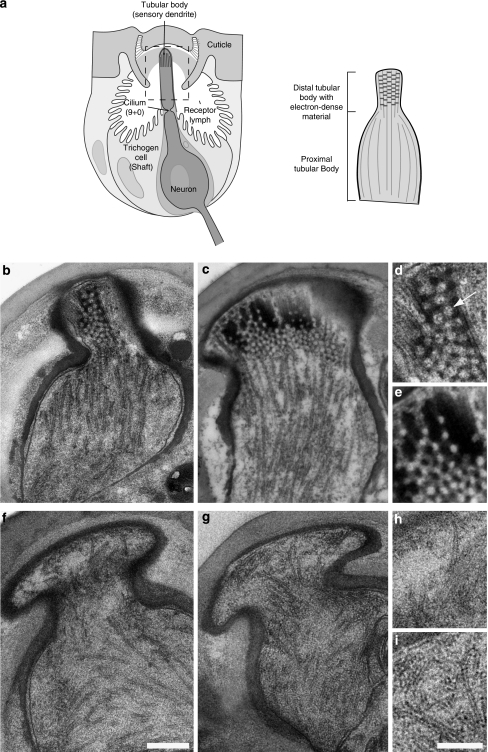
Figure 4. The ultrastructure of the tubular body in f02655 campaniform receptors in the Drosophila haltere is altered.
(a) Schematic drawing of the campaniform receptor. The tubular body of the receptor is marked and illustrated separately (right panel). (b, c) Electron micrographs of the sensory dendrite in wild-type campaniform receptors in the haltere. The distal part of the tubular body shows characteristic electron-dense material. (d, e) Electron-dense material in the distal tubular body at higher magnification. (f, g) Electron micrographs of f02655 campaniform receptors. The electron-dense material in the distal tubular body is missing. Microtubules in the proximal tubular part seem disorganized. (h, i) Distal tubular bodies of f02655 campaniform receptors at higher magnification. Scale bars represent 0.5 μm (f) and 250 nm (i).

