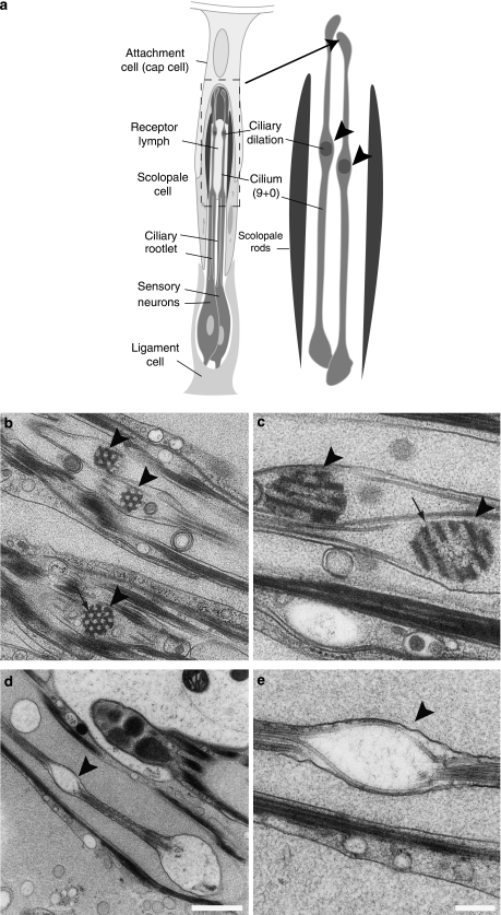
Figure 5. Mutant f02655 sensory neurons of Johnston's organ display an altered ultrastructure.
(a) Schematic representation of Johnston's organ in sensory neurons of Drosophila antenna. The region with the ciliary dilation is marked and illustrated separately (right part). (b, c) Electron micrographs of ciliary dilations (arrowheads) in wild-type chordotonal neurons with electron-dense material in the ciliary dilation (arrows point towards regular holes and channels in the electron-dense material). (d, e) Chordotonal neuron of f02655 flies with 'empty' ciliary dilations. Note how the microtubules are still bent as in wild-type dilations. Scale bars represent 1 μm (d) and 250 nm (e), arrowheads point towards ciliary dilation.

