Figure 2.
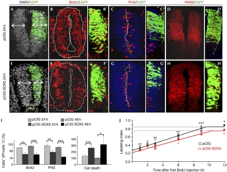
Forced SOX5 expression promotes cell cycle exit. In relation to control pCIG embryos (A–C′), SOX5 misexpression (pCIG-SOX5; GFP, green on right side; E–H′) in HH14–16 embryos caused a 30% reduction in the size of the hemitube (E), in the number of BrdU (F,F′) and PH3-positive cells (G,G′), and an increase in activated caspase 3 (Cas3*)-positive dying cells (I). Expression of the neural progenitor marker PAX6 is not altered in SOX5HIGH cells (H,H′, D,D′). (I) Quantification of the effect 24 or 48 h PE. *P<0.01; **P<0.005; ***P<0.001. (J) Cumulative BrdU labelling curves of pCIG (black squares) or pCIG-SOX5 (red circles) electroporated neural tube cells. Dashed lines indicate the reduction in growth fractions. Mean of three embryos per experimental point, s.d. and t-test was calculated; ***P<0.01; **P<0.025; *P<0.05. BrdU, bromodeoxyuridine; C, control; EP, electroporated; GFP, green fluorescent protein; HH, Hamburguer and Hamilton; pCIG, pCAGGS-IRES-GFP; PE, post-electroporation; PH3, phospho-histone H3.

