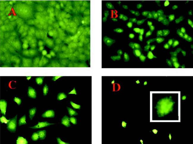
Fig. 4.
Acridine orange staining of A549 cells: A, untreated control cells; B, cells exposed to aerosolized nimesulide (40 shots); C, cells treated with doxorubicin (0.25 μg/ml); D, cells treated with aerosolized nimesulide (40 shots) + doxorubicin (0.25 μg/ml). Original magnification 40×. Inset to D, 100× magnification of an apoptotic cell. The cells were exposed to aerosolized nimesulide on the fifth stage of the viable impactor and incubated for 15 min, and 1 ml of cell suspension was transferred to chamber slide. The slide was incubated for 72 h and stained with acridine orange as described in the Materials and Methods.

