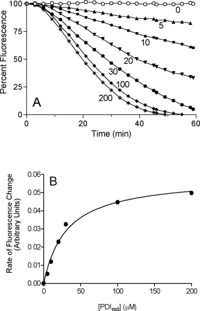
FIGURE 6.
The effect of increasing concentrations of reduced PDI on the refolding of RfBP driven by QSOX. Panel A: the conditions of Figure 5 were used except that a concentration of 30 nM QSOX was used and the reduced PDI concentration was varied from 0 to 200 μM. Panel B: the maximal rates of riboflavin rebinding attained for each curve in panel A show a hyperbolic dependence with half-saturation at 30 μM reduced PDI.

