
Figure 5.
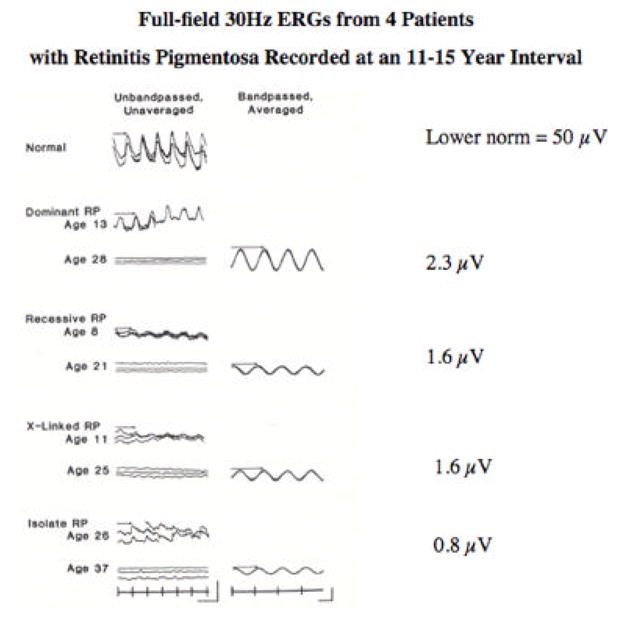
Full-field 30-Hz cone ERGs from a normal subject and four patients with retinitis pigmentosa tested at an 11–15-year interval. Stimulus onset, vertical markers; calibration symbol (left column, lower right) designates 100 μV vertically for the normal subject and top three patients and 40 μV vertically for the bottom patient and 50 msec horizontally for all traces; calibration (right column, lower right) designates 2 μV vertically for the dominant, X-linked, and isolate patients and 0.3 μV for the recessive patient and 20 msec horizontally for all traces. B-wave implicit times are designated with arrows. (From Andreasson, et al., 1988.)