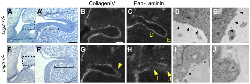
Fig. 4. The basement membrane undergoes early and ectopic breakdown in the inner ear of Lrig3 mutant mice.
(A,F) Transverse plastic sections through inner ears of E12 Lrig3 +/− (A) and −/− littermates (F), with magnified views of the boxed areas (A′, F′). Dorsal is up; lateral is to the right. In controls, epithelial cells in the fusion plate intercalate to form a single layer of cells (A′), but in mutants, this region is expanded (brackets, F′). Scale bar: 50 μm. (B, C, G, H) Immunofluorescent detection of Collagen IV (B,G) and all laminins (C, H) in E12 +/− (B,C) and −/− embryos (G,H) sectioned in the transverse plane. In homozygotes, the basal lamina is disturbed by breaks in the laminin network and ectopic accumulation of Collagen IV (arrowheads, G, H). (D, E, I, J) Electron micrographs of the regions indicated in C and H. The basement membrane is continuous in heterozygotes (arrowheads, D, E) but is absent (asterisks, I) or severely disrupted (asterisk, J) in homozygotes. Scale bar: 500 nm.

