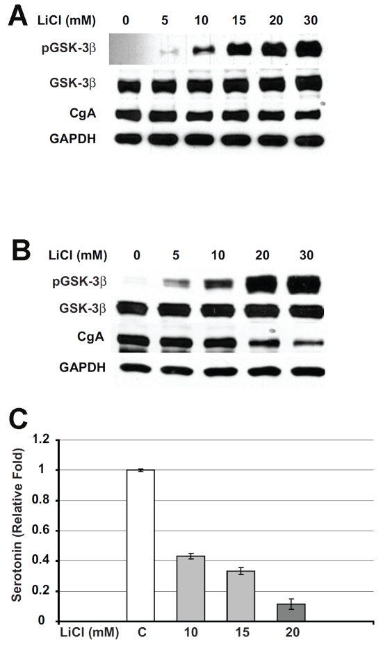
Figure 2.
Lithium treatment leads to GSK-3β phos-phorylation and suppression of neuroendocrine tumor markers in carcinoid cells. Carcinoid cells were treated with the indicated concentrations of lithium chloride (LiCl) for 2 days and immunoblotting was performed on whole cell lysates for phosphorylated GSK-3β, total GSK-3β, and the neuroendocrine markers chromogranin A (CgA) and serotonin. A. LiCl treatment increased the level of phosphorylated GSK-3β, whereas total GSK-3β was relatively unchanged in BON cells. GAPDH was used as a loading control. B. Total cellular extracts from H727 cells treated with LiCl were analyzed by Western blot for the levels of phosphorylated GSK-3β and neuroendocrine markers. LiCl treatment led to an increase in phosphorylated GSK-3β protein. Note that in control cells there was no detectable level of phosphorylated GSK-3β. In contrast to this, regular active GSK -3β was present at high levels in both control and treated cells with no change in the levels in treated cells. The increase in the levels of phosphorylated GSK-3β was associated with a significant reduction in CgA. GAPDH was used to confirm equal protein loading. C. Serotonin levels were measured by ELISA in LiCl-treated BON carcinoid cell lysates. Lithium treatment dose-dependently reduced the levels of serotonin in BON cells.

