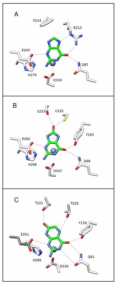
Figure 8.
Models for substrate binding in the active sites of guanine deaminase, 8-oxoguanine deaminase and isoxanthopterin. (A) Guanine was placed in the active site of human guanine deaminase (PDB code: 2UZ9) via a simple substitution of this compound for the product xanthine. (B) 8-Oxoguanine was positioned in the active site of Sgx9236e (PDB code: 3HPA) using a structural overlay of this protein with the active site of guanine deaminase (PDB code: 2UZ9) while overlapping the common atoms of 8-oxoguanine and xanthine. (C) Isoxanthopterin was positioned in the active site of Sgx9339a using a structural overlay of this protein with the active site of guanine deaminase while overlapping the common atoms of isoxanthopterin and xanthine. Since residues 251-263 are disordered in 2PAJ, the important glutamate (Glu-251) from the HxxE motif at the C-terminal end of β-strand 5 was inserted into the active site based upon the orientation of Glu-243 in guanine deaminase (PDB code: 2UZ9).

