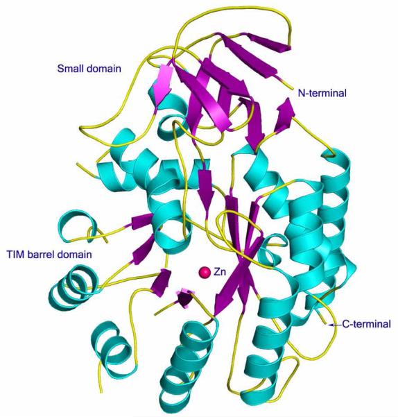
Figure 2.
Ribbon representation of the overall structure of Sgx9339a (PDB code: 2PAJ). The α-helices, β-strands and random coil loops are colored cyan, violet and yellow, respectively. The zinc ion is colored in pink. The small domain is made up of the N-terminal residues 10-70 and of residues 413-456. The ribbon diagram was made using PYMOL.

