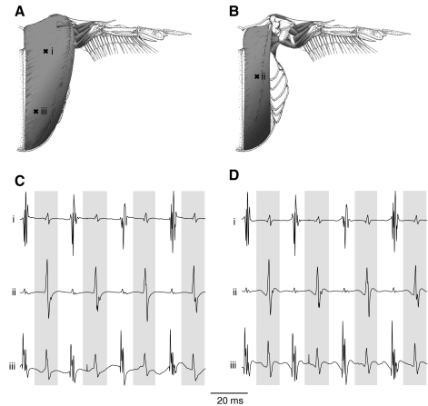
Fig. 2.
Representative electromyograms (EMGs) from the pectoralis major and supracoracoideus of an individual Anna's hummingbird from a preliminary experiment. Electrodes i and iii were placed in the left pectoralis major (A), and electrode ii was placed in the underlying supracoracoideus (B) at the sites indicated by ‘x’. Hummingbird musculoskeletal drawings were adapted from Welch and Altshuler (Welch and Altshuler, 2009). Representative traces from the three EMG electrodes are presented as direct output from the amplifier with filter cut-offs of 1 Hz and 10 kHz (C). Upstrokes are indicated by the white spaces in between the gray bars, which indicate the downstrokes. The same traces are presented following offline filtering (D). The time scale applies to both raw amplifier output and post-processed signals.

