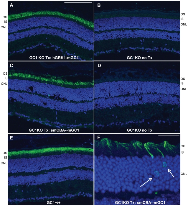
Figure 1. AAV-mediated GC1 expression in photoreceptors of the GC1KO mouse.
AAV5-hGRK1-mGC1 drives expression of GC1 in photoreceptor outer segments of GC1KO mice. (A). No GC1 expression is seen in the untreated, contralateral control eye (B). AAV5-smCBA-mGC1 drives expression of GC1 in photoreceptor outer segments (C) and occasionally in photoreceptor cell bodies (arrows in F). No such GC1 expression is seen in the untreated, contralateral control eye (D). Levels of therapeutic transgene expression in the AAV5-mGC1-treated eyes are only slightly less than that seen in isogenic GC1+/+ control eyes (E). All retinas were taken from mice 3 months post treatment or age matched untreated controls. Scale bars in A = 100µm, F = 25µm. OS-outer segments, IS-inner segments, ONL-outer nuclear layer.

