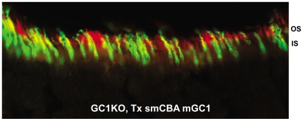
Figure 2. AAV5-mGC1 drives expression of GC1 in both rod and cone photoreceptors.
Representative retinal section from a GC1KO eye injected with AAV5-smCBA-mGC1 stained for GC1 (red) and PNA lectin (green) reveals GC1 expression in cone outer segments (yellow overlay) as well as in rod outer segments (red alone). hGRK1-mGC1 injected eyes revealed the same pattern (data not shown).

