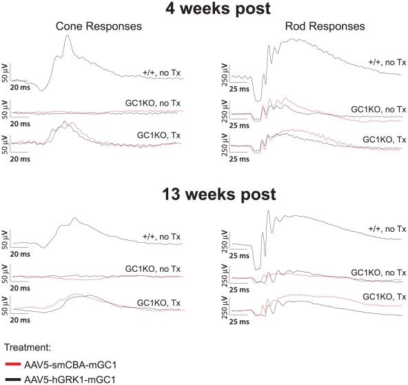
Figure 3. AAV5-mGC1 restores retinal function to cone photoreceptors of the GC1KO mouse.
Representative cone (left column)- and rod (right column)-mediated ERG traces from GC1 +/+ (upper waveforms), untreated GC1KO (middle waveforms) and AAV5-mGC1-treated (bottom waveforms) mice. For the middle and bottom waveforms in each panel, red traces correspond to eyes injected with AAV5-smCBA-mGC1 (bottom) and their uninjected contralateral eyes (middle) and black traces correspond to eyes injected with AAV5-hGRK1-mGC1 (bottom) and uninjected contralateral eyes (middle). Cone responses in AAV5-mGC1-treated eyes are restored to approximately 45% of normal amplitude.

