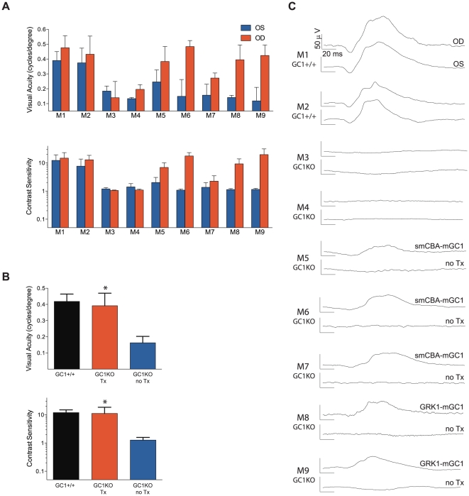
Figure 5. Optomotor analysis of visual function restoration in GC1KO mice treated with either AAV5-smCBA-mGC1 or AAV5-hGRK1-mGC1.
M1–M9 correspond to the nine mice used for testing. Photopic acuities and contrast sensitivities of GC1+/+ control mice (M1, M2), naïve GC1KO (M3, M4), smCBA-mGC1-treated (M5, M6, M7) and hGRK1-mGC1-treated GC1KO (M8, M9) mice reveal that treated mice behave like normal sighted mice (A, B). Average values for photopic visual acuity and contrast sensitivity of all GC1+/+ eyes (n = 4), untreated GC1KO eyes (n = 9) and AAV5-mGC1-treated eyes (n = 5) are shown (B) (* = P<0.0001). Cone-mediated ERG responses from each mouse (M1–M9) are shown for comparison (C).

