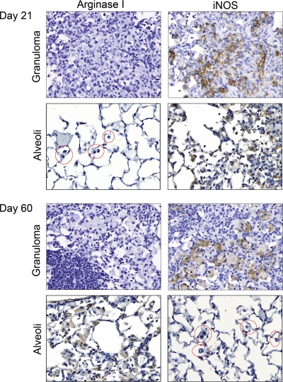
Figure 5.
IHC staining of granulomas for iNOS and arginase I. IHC analysis of arginase I and iNOS in M. tuberculosis infected lungs 21 and 60 days after infection. Alveolar macrophages are circled in red. Granuloma-associated macrophages and alveolar macrophages exhibit iNOS staining starting 21 days after infection. By Day 60, alveolar macrophages express arginase I and not iNOS. Alveolar and granuloma macrophage IHC staining was photographed in the same lung at each time-point, and alveolar staining was examined near and distal to granulomas and found to be similarly stained for arginase I and iNOS regardless of distance from the granulomas. Final original magnification, ×400.

