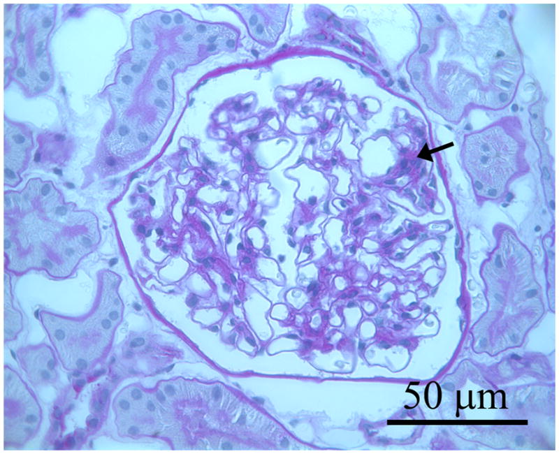
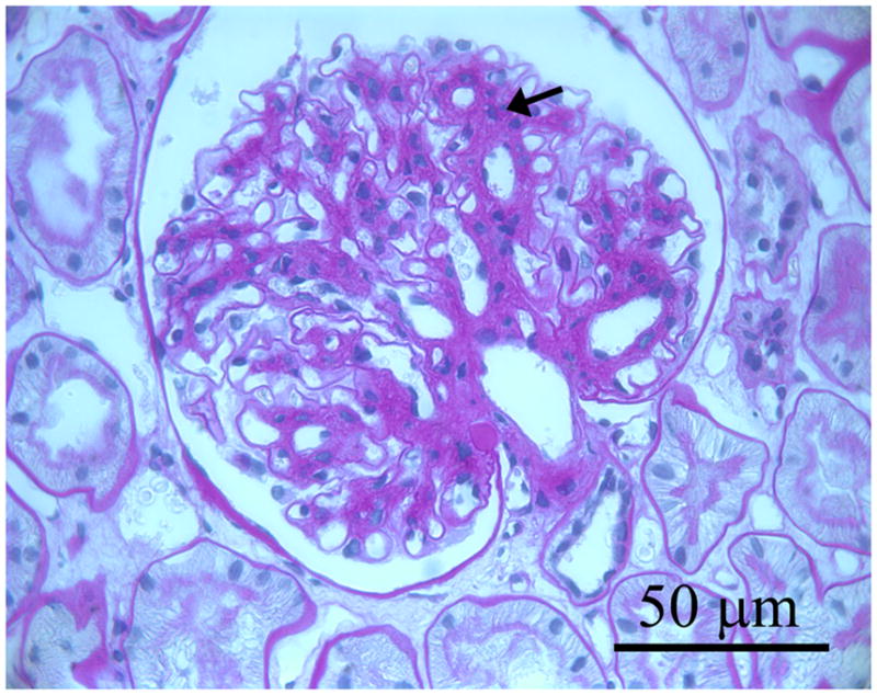
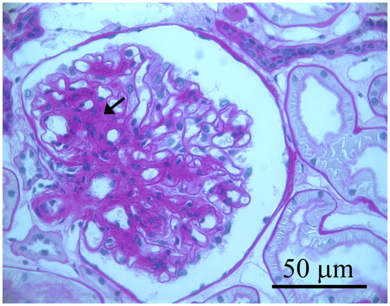
Figure 1.


Three photomicrographs from three separate baseline biopsies of glomerular tissues for light microscopy that were fixed in Zenker’s solution and embedded in paraffin and stained with periodic acid Schiff (PAS): a. glomerulus in which the mesangium appears normal; b. shows a glomerulus with mild mesangial expansion with increased PAS positive matrix material between the capillary loops and branching from the base of the glomerulus (hilus) to the periphery (black arrows); c. shows a glomerulus with moderate mesangial expansion (black arrows).
