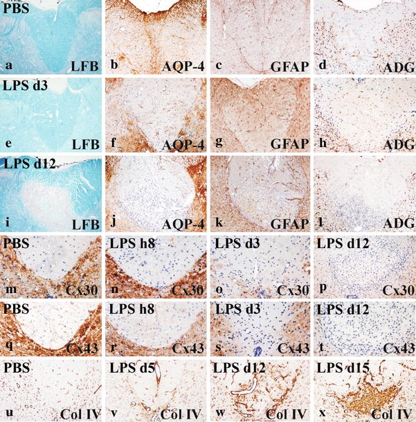
Fig. 2.

Astrocyte pathology and demyelination in LPS-induced spinal cord lesions: a–d no pathological alterations of astrocytes and no demyelination are seen in animals injected with saline into the spinal cord white matter. 3 days after injection of LPS, there is no demyelination (Luxol fast blue staining e); staining for aquaporin 4 is reduced (f), but astrocytes are still preserved (g). In contrast, there is a profound loss of α-dystroglycan reactivity at the vascular glia limitans (h). At day 12 after LPS injection, a focal plaque of demyelination is visible (Luxol fast blue staining (i), associated with complete loss of aquaporin 4 (j) and α-dystroglycan reactivity (l) and a profound reduction in astrocytes and their processes (k). Connexin 30 immunoreactivity is progressively lost with time in LPS lesions (n–p) in comparison with saline-injected animals (m). q–t Connexin 43 immunoreactivity shows similar changes in comparison with that for connexin 30. u–x LPS lesions show an increasing amount of collagen IV deposited in the spinal cord extracellular space with time. Bar 100 µm
