
Fig. 4.
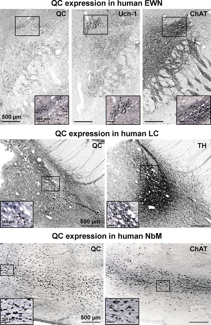
QC expression in the human Edinger–Westphal nucleus, locus coeruleus and nucleus basalis Meynert. QC immunoreactivity was present in midbrain sections in a defined cell group (top left). These cells include both Ucn-1 and ChAT expressing subpopulations of the Edinger–Westphal nucleus as shown by the labelling of these marker proteins in adjacent brain sections. QC-immunoreactive neurons were also detected in brainstem sections in a small, defined cell group within the locus coeruleus area (middle left). Labelling of consecutive brain sections with TH identified these neurons as noradrenergic LC neurons (middle right). The highest density of QC-immunoreactive neurons was detected in the nucleus basalis Meynert (bottom left), which was identified by the presence of cholinergic Ch4 neurons (bottom right). Scale bars in overview 500 μm, in higher magnification inserts 100 μm, 150 μm and 200 μm as indicated