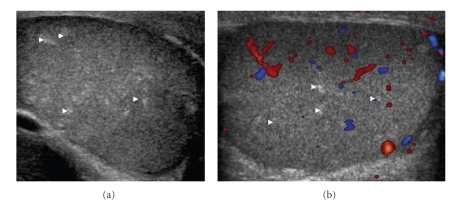
Figure 1.
26-year-old patient presented with painless swelling in the right scrotum. (a) Grey Scale Ultrasound of Scrotum showing multiple small ill defined and randomly distributed echogenic foci within the testicular substances (white arrowheads). (b) Colour Doppler showing normal vascularity of testis. However, the lesions are typically avascular.

