
Figure 4. Architecture of the VSV virion and implication for pseudotyping.
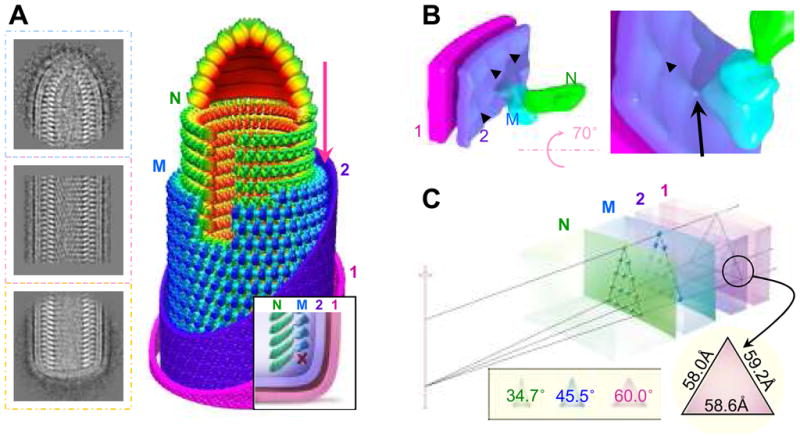
(A) Representative 2D averages of conical tip, trunk and base of VSV and a montage model of the tip and the cryoEM map of the trunk. N is green, M is blue, and the inner (“2”) and outer (“1”) leaflets of the membrane are violet and pink. Inset: illustration of the base region of the VSV virion. The “X” marks the absence of a turn of M helix below the lowest turn of the N helix. (B) CryoEM structure showing the putative cytoplasmic tail of G protein binding to an M subunit through a thin linker. The inner leaflet (“2”) of the membrane has unusual bumps (arrowhead) that meet M at the site of a thin linker density (arrow). (C) A wedge of the virion trunk, illustrating its geometric arrangement across the three layers. The N layer, the M layer, and the two membrane leaflets (“1” and “2”) are arranged in coaxial cylinders with their radii determined from our cryoEM structure. Due to the difference in radii, the helical lattice points on the three layers form different triangles. The smaller the radius, the narrower the apex angle (inset). At the outer surface of the membrane, the lattice points form an equilateral triangle.