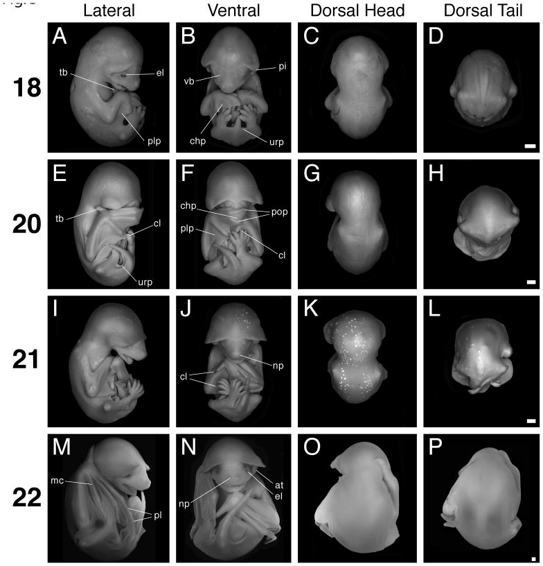
Fig. 3.
Stages 18 to 22. Scale bar = 1 mm in all panels. The first column (A, E, I, M) shows lateral views with dorsal to the left, the second column (B, F, J, N) shows ventral views, the third column (C, G, K, O) shows dorsal views of head and trunk, and the fourth column (D, H, L, P) shows dorsal views of the trunk and tail. A–D: Stage 18 specimen. E–H: Stage 20 specimen. I–L: Stage 21 specimen. M–P: Stage 22 specimen. at, antitragus; chp, chiropatagium; cl, claw; el, eyelid; mc, metacarpal; np, nasal pit; pi, pinna; pl, phalanx; plp, plagiopatagium; pop, posterior oriented phalanx; tb, thumb; urp, uropatagium; vb, developing vibrissa.

