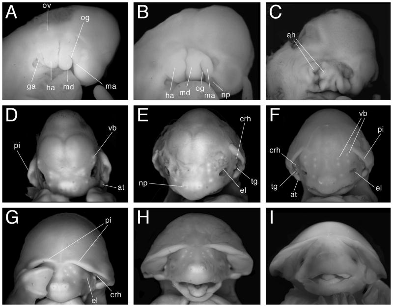
Fig 5.
Craniofacial development. Panels A–C show lateral views of developing head with anterior to the right. Panels D–I show face-on views with anterior to the top. A. Stage 13. B. Stage 14. C. Stage 15. D. Stage 16. E. Stage 17. F. Stage 18. G. Stage 20. H. Stage 21. I. Stage 22. ah, auditory hillock; at, antitragus; crh, crus helix; el, eyelid; ga, glossopharyngeal arch; ha, hyoid arch; ma, maxilla; md, mandible; np, nasal pit; og, oral groove; ov, otic vesicle; pi, pinna; tg, tragus; vb, developing vibrissa. Views are not to scale.

