
Fig. 3.
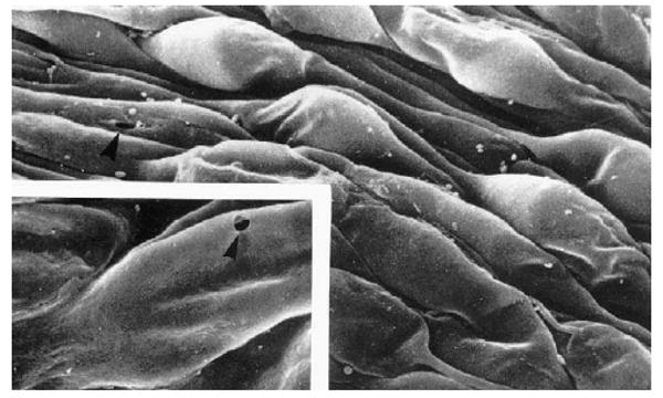
Scanning electron micrograph of the inner wall of Schlemm's canal as viewed from within the canal. The bulging structures are giant vacuoles (and some nuclei). The insert shows an arrowhead pointing at a pore passing through one of the giant vacuoles (modified from (Allingham et al., 1992)). © 1992, Association for Research in Vision and Ophthalmology.