
Fig. 4.
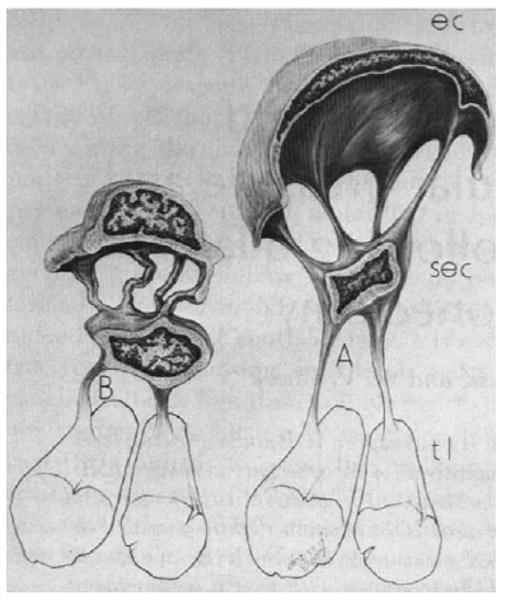
Schematic diagram showing change in configuration of cell (EC) of endothelial lining of Schlemm's canal going from low IOP (B) to high IOP (A). These cells attach to a second layer of cells (SEC) that in turn attach to the trabecular lamella (tl). (Johnstone, 1979). © 1979, Association for Research in Vision and Ophthalmology.