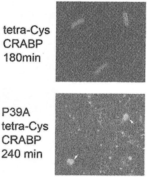
Fig. 7.3.
Fluorescence microscopy images showing uniformly distributed fluorescence of tetra-Cys CRABP (180 min after induction) and hyperfluorescent dense aggregates of P39A tetra-Cys CRABP at the poles of the cells (at 240 min after induction). Hyperfluorescent impurities in the extracellular medium are marked by an arrow.

