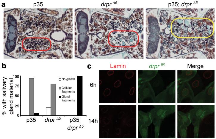
Figure 2. Draper functions downstream or in parallel to caspases during salivary gland cell death.
a, Animals with salivary gland-specific expression of p35 (fkh-GAL4/+; UAS-p35/+), n=18, drpr null animals (+/w; +/UAS-p35; drprΔ5/drprΔ5), n=10, and drpr null animals with salivary gland-specific expression of p35 (fkh-GAL4/w; UAS-p35/+; drprΔ5/drprΔ5), n=16, were analyzed by histology for the presence of salivary gland material 24h after puparium formation. Cell fragments are in red circles, and gland fragments are in the yellow circle. b, quantification of data from a. c, Salivary glands were dissected from animals expressing drprIR specifically in GFP-marked clone cells (hsflp/w; UAS-drprIR/+; act<FRT,cd2, FRT>Gal4, UAS-GFP/+) 6h and 14h after puparium formation. Salivary glands were stained with GFP antibody (green) to label cells expressing drprIR, and Lamin antibody (red).

