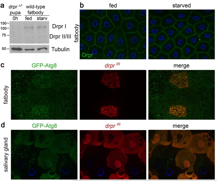
Figure 4. Drpr is cell-autonomously required for autophagy in dying salivary glands, but not in response to starvation in the fatbody.
a, Protein extracts from drpr null (w; drprΔ5/drprΔ5) pupae at puparium formation (0h) and from the fatbodies of wild type (Canton-S) third instar larvae were analyzed by Western Blotting with anti-Drpr antibody. Third instar larvae were either fed or starved 4h. b, Wild-type (Canton-S) third instar larvae were either fed or starved 4h, and fatbodies were dissected, stained with anti-Drpr antibody, and imaged for Drpr (green). Nuclei were stained with DAPI (blue). c, Third instar larvae expressing GFP-Atg8 in all cells, and drprIR specifically in dsRed-marked clone cells (hsflp/w; UAS-drprIR/+; hsGFPAtg8b, act<FRT,cd2, FRT>Gal4, UAS-dsRed/+), were starved 4h. Larval fatbodies were dissected and imaged for GFPAtg8 (green) and dsRed (red). d, Salivary glands of animals expressing GFP-Atg8 in all cells, and drprIR specifically in dsRed-marked clone cells (hsflp/w; UAS-drprIR/+; hsGFPAtg8b, act<FRT,cd2, FRT>Gal4, UAS-dsRed/+) were dissected 14h after puparium formation. Salivary glands were imaged for GFPAtg8 (green) and dsRed (red). Nuclei were stained with Höescht (blue).

