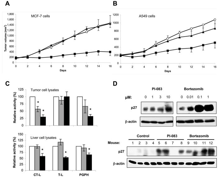
Figure 6. Effects of PI-083 and Bortezomib on tumor growth and the levels of the proteasomal substrate p27kip1 in vivo.
The growth of human tumors following the injection of (A) MCF-7 breast cancer and (B) A549 lung cancer cells into nude mice was determined as described in Materials and Methods. Mice were treated with DMSO (○), 1 mpk Bortezomib (▲) or 1 mpk PI-083 (□). Data represent the means ± standard error of one of three independent experiments with 4 to 6 animals in each group. Asterisks indicate statistical significance (p < 0.05). Proteasomal activities in tumor cell lysates (C, upper panel) or liver cell lysates (C, lower panel) following treatment with DMSO (n=4, white), PI-083 (n=4, gray) or Bortezomib (n=4, black). The asterisks indicate p values ≤ 0.006 for a comparison of experimental and DMSO-treated mice. (D, upper panel) A549 lung cancer cells were treated with 0.1 % DMSO (lane 1) or water (lane 5) or the indicated drug concentrations for 48 h. Cell lysates were then subjected to Western blot analyses with antibodies to p27Kip1 and β-actin as a loading control. (D, lower panel) p27Kip1 levels were determined in lysates prepared from vehicle- or drug-treated A549-tumors by Western blots, with β-actin serving as a loading control.

