
Fig. 4.
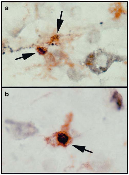
The VDR protein expression in human brains. VDR was detected in microglia in neocortical tissues using double immunohistochemistry of a VDR antibody (Epitomics) and the MHCII antibody (LN3). VDR immunoreactivity is in black and the MHCII immunoreactivity is in brown (indicated by arrows). VDR immunoreactivities exhibit in granular profiles (a), and intracellular profile resembling nuclear localization (b)