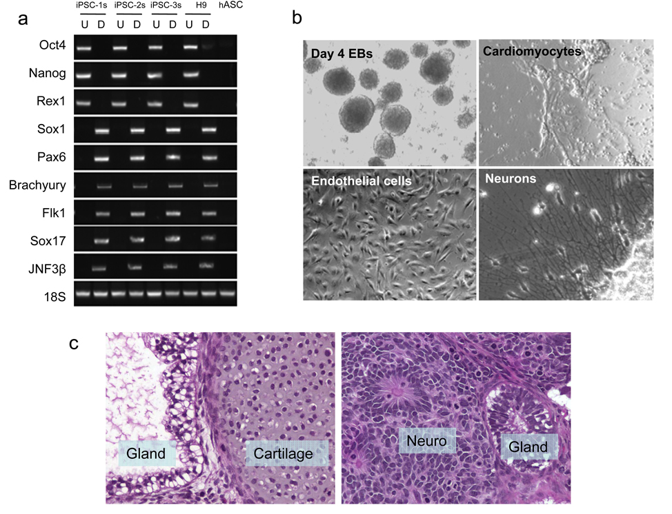
Figure 2. Pluripotency of mc-iPS cells.
a, RT-PCR analysis of pluripotent markers (Oct4, Nanog and Rex1) and various differentiation markers for the three germ layers (ectoderm: Sox1, Pax6; mesoderm: Brachyury, Flk1; endoderm: Sox17, JNF3β). Undifferentiated (U) mc-iPS cells; differentiated (D) mc-iPS cells after 8 days suspension culture followed by 8 days adherent culture. b, Multiple cell types were differentiated from mc-iPS cells. See Supplementary Figure 9 and Supplementary Video 1 for further characterization. c, Subcutaneous injection of mc-iPS cells causes teratomas in SCID mice consisting of all three embryonic germ layers, including epithelial cells (ectoderm), cartilage (mesoderm), and glandular structures (endoderm). Tissue sections from subclone mc-iPSC-1s are shown as representative.

