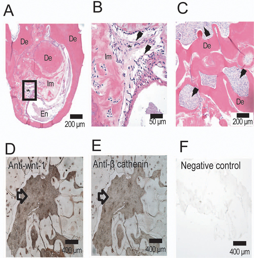
Figure 2.
Cellular components of odontoma. Hematoxylin-eosin-stained section of odontoma (A) showing outer layer of dentin (De) surrounding inner layers of enamel (En), immature matrix (Im) and a rich cellular components (*). Higher magnification of cellular components in A (box) is presented in B. This shows heterogeneous staining fibroblast-like cells (arrows) mixed with immature matrix at different stages of mineralization (B). Another section of the odontoma showed compartmentalization of the cellular fibroblastic network by dentin-like matrix (C). Odontoma cellular contents immunoreacted positively with goat polyclonal antibody to human Wnt1-1 (D) and mouse monoclonal antibody to human β-catenin (E). Non-immune goat serum (F) and IgG isotype control were unreactive.

