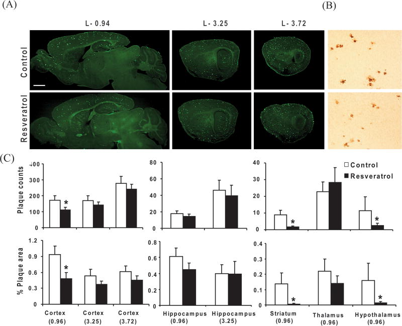
Fig. 3. Resveratrol and plaque pathology.
Forty five days old Tg19959 mice were fed AIN 93G diet with or without 0.2% resveratrol. Animals were sacrificed at 90 days old, and the brains were processed for thioflavine S staining. (A) Representative saggital brain sections from lateral levels − 0.94, − 3.25, − 3.72, respectively, showed that resveratrol decreased thioflavin S immunoreactivity (Scale bar, 500 μm). (B) Resveratrol also increased decreased 6E10 immunoreactivity stained with 6E10 antibody directed against 1–17 amino acids of Aβ in representative cortex sections (Scale bar, 200 μm). (C) Graphs shows the plaque counts and % area occupied by plaques quantified from the cortex, hippocampus, striatum, thalamus and hypothalamus region. Data represent means ± SEM of control (n=8) and 0.2% resveratrol (n=9) groups from 2–3 independent experiments. * Denotes control differs from resveratrol group (p<0.05).

