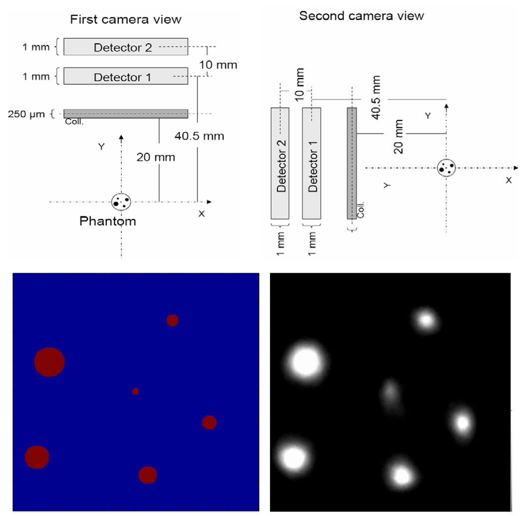
Figure 14.
Simulation study with SiliSPECT: The projections from the phantom were acquired at two different magnifications, M1 = 1, M2 = 1.5 (top). The phantom consists of microstructure capillary tubes of diameters between 60 and 250 µm. A slice from simulated data reconstructed with MLEM algorithm is shown in the right figure below.

