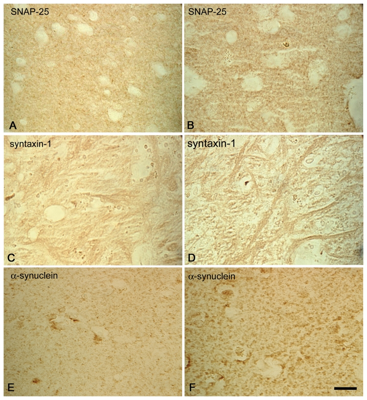
Figure 7.
Redistribution of SNARE proteins in Parkinson’s disease. Immunohistochemistry for SNAP-25 (A, B), syntaxin-1 (C, D) and α-synuclein (E, F) in the striatum (putamen A, B, E, F; external globus pallidus C, D) of control (A, C, E) and Parkinson’s disease (B, D, F) brains. Images are representative of n = 3 for control brains and n = 4 for Parkinson’s disease brains. Scale bar = 5 µm.

