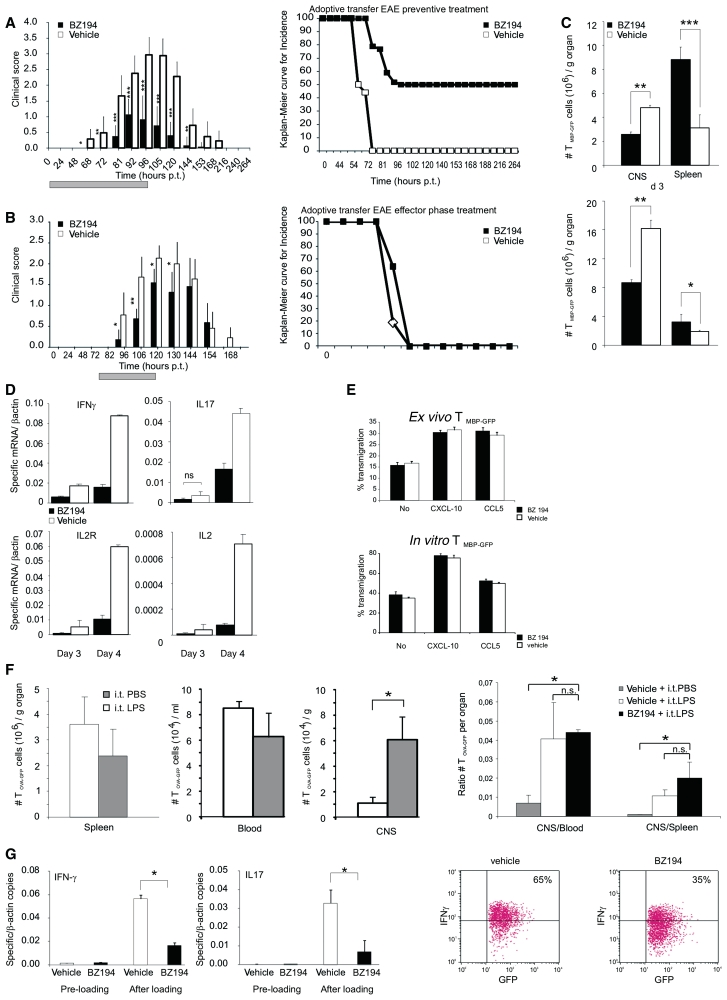
Figure 4.
NAADP-antagonist interferes with TMBP-GFP cell activation within the CNS. (A and B) Treatment of adoptive transfer EAE. Clinical course (left) and disease incidence (right; Kaplan–Meier curve) of EAE after transfer of TMBP-GFP cells. Treatment performed with preventive schedule (A; n = 22 animals/group) and during effector phase (B; n = 8 animals/group) with BZ194 (black) or vehicle (DMSO, white). Grey bar: period of treatment: 0–96 h post transfer (p.t.) (A); 72–120 h p.t. (B). Cumulative data of five (A) and three (B) independent experiments are shown (mean values ± SD). P-values measured by Mann–Whitney rank statistical test: *P ≤ 0.01, **P ≤ 0.001, ***P ≤ 0.0001. (C) BZ194 reduces homing of encephalitogenic effector T cells into their target organ. Cytofluorometric quantification of TMBP-GFP cells in the spinal cord (CNS) or the spleen of BZ194-treated (black) or vehicle-treated (white) animals 3 days (upper panel) and 4 days (lower panel) p.t. Values represent mean values ± SD of triplicate measurements. Representative data of five independent experiments. P-values: *P ≤ 0.05, **P ≤ 0.01, ***P ≤ 0.005. (D) NAADP signalling is essential for efficient re-activation of effector TMBP-GFP cells within their target organ. Reduced expression of pro-inflammatory mediators in autoaggressive TMBP-GFP cells within the CNS after BZ194 treatment. Quantitative PCR of TMBP-GFP cells sorted from spinal cords 3 and 4 days p.t. Black bars: BZ194-treated; white bars: vehicle-treated animals. Values are normalized to β-actin mRNA. Representative data of three independent experiments. All differences are statistically significant (P-value at least ≤0.05) unless otherwise specified. ns, not significant. (E and F) BZ194 treatment does not interfere with migration capacity of ex vivo, in vitro or in vivo effector T cells. (E) TMBP-GFP cells were isolated ex vivo from spleens (upper plots) of animals treated with BZ194 (0.5 mM) (black bars) or vehicle (white bars) at Day 3 p.t., or cultured (lower plots) for 3 days in presence of the inhibitor (black bars) or vehicle (white bars). Percentage of transmigration without or in presence of the indicated chemokines was assessed by cytofluorometry 6 h after plating. (F) Three days after transfer of 2.5 × 10⁶ effector TOVA-GFP cells, animals were intrathecally (i.t.) injected with 150 ng of lipopolysaccharide (LPS) (white bars) or equal volume of phosphate buffered saline (PBS) (grey bars); 24 h later, the numbers of GFP positive cells were determined in the spleen (left plot), blood and spinal cord (CNS; middle plot) by flow cytometry. Right plot: adoptive transfer of effector TOVA-GFP cells was performed as above. The animals received a daily i.p. injection of BZ194 or vehicle from Days 0 to 3 p.t. At the Day 3 p.t. BZ194-treated (black bars) and vehicle-treated (white bars) animals were i.t. injected with lipopolysaccharide; for control, vehicle-treated animals were also i.t. injected with phosphate buffered saline (grey bars). The graph shows ratios of total number of GFP-positive cells detected in the spinal cord (CNS) relative to the numbers in the blood (left) or in the spleen (right) on Day 4 p.t. *P < 0.05; n.s. = not significant. (G) BZ194 treatment reduces the activation of effector T cells on CNS explants. TMBP-GFP cells were cultured for 2 h in presence of BZ194 (black bars, pre-loading) or vehicle (white bars, pre-loading) and then loaded on spinal cord explants from mild EAE animals (after loading) in presence of the antagonist (0.5 mM; black bars) or of the vehicle (white bars). Twenty-four hours later, T MBP-GFP cells were sorted out and the levels of IFNγ (left graph) and interleukin-17 (right graph) were measured by quantitative polymerase chain reaction. Representative data of three independent experiments are shown. Values normalized to β-actin mRNA. *P < 0.05. Right dot plots: corresponding protein expression of IFN-γ, evaluated by flow cytometry of TMBP-GFP cells isolated from spinal cord explants 36 h after loading.

