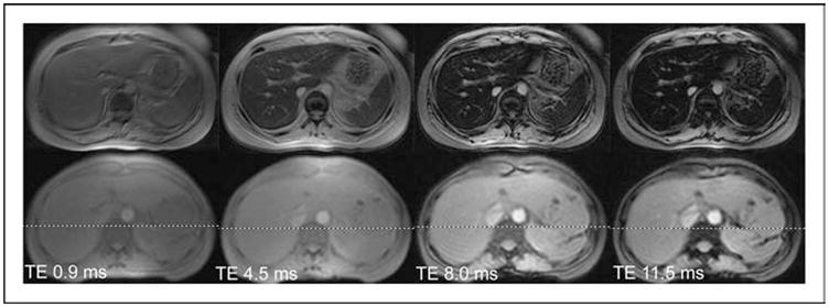
Figure 1. Gradient echo images of liver collected at four different echo times.

The top four images were collected from a patient having a liver iron of 6 mg/g. The bottom four images were collected from a normal volunteer. All images darken as the echo time (TE) lengthens, but the iron-heavy tissue darkens faster. The half life of this process is called T2* and the rate is called R2* (R2*=1000/T2*).
