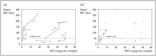
Figure 5. Iron trajectories of five patients.
(a) Scattergram demonstrating cardiac iron versus liver iron (HIC) in three patients. Temporal trajectories trace out an oval, demonstrating the lack of cross-sectional correlation between liver and heart iron. (b) Same scattergram with two additional patients who developed cardiac iron loading despite having apparently adequate iron chelation.

